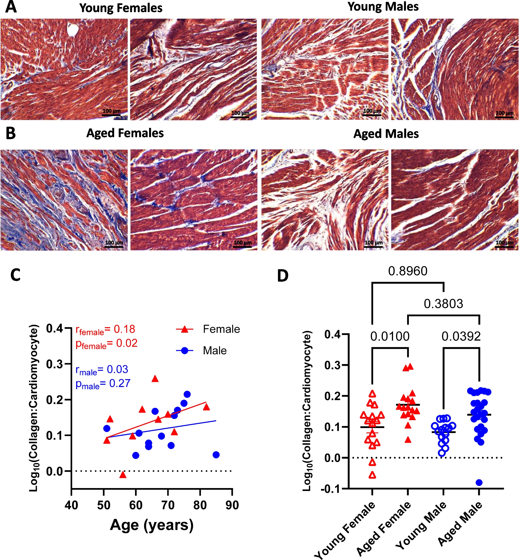
United Nations. World social report 2023: leaving no one behind in an ageing world. 2023.
Kim-Dorner SJ, et al. Age- and gender-based comorbidity categories in general practitioner and pulmonology patients with COPD. NPJ Prim Care Respir Med. 2022;32(1):17.
Article
Google Scholar
Jaul E, Barron J. Age-Related Diseases and Clinical and Public Health Implications for the 85 Years Old and Over Population. Front Public Health. 2017;5:335.
Article
Google Scholar
Rodgers JL, et al. Cardiovascular Risks Associated with Gender and Aging. J Cardiovasc Dev Dis. 2019;6(2):19.
CAS
Google Scholar
Oneglia A, Nelson MD, Merz CNB. Sex Differences in Cardiovascular Aging and Heart Failure. Curr Heart Fail Rep. 2020;17(6):409–23.
Article
Google Scholar
Keller KM, Howlett SE. Sex Differences in the Biology and Pathology of the Aging Heart. Can J Cardiol. 2016;32(9):1065–73.
Article
Google Scholar
Daniel RG, Ahmed A-L. Immune mechanisms of cardiac aging. J Cardiovasc Aging. 2023;3(2):17.
Google Scholar
Biernacka A, Frangogiannis NG. Aging and Cardiac Fibrosis. Aging Dis. 2011;2(2):158–73.
Google Scholar
Lu L, et al. Cardiac fibrosis in the ageing heart: Contributors and mechanisms. Clin Exp Pharmacol Physiol. 2017;44(Suppl 1):55–63.
Article
CAS
Google Scholar
Travers JG, et al. Cardiac Fibrosis: The Fibroblast Awakens. Circ Res. 2016;118(6):1021–40.
Article
CAS
Google Scholar
Martos R, et al. Diastolic heart failure: evidence of increased myocardial collagen turnover linked to diastolic dysfunction. Circulation. 2007;115(7):888–95.
Article
Google Scholar
Reed AL, et al. Diastolic dysfunction is associated with cardiac fibrosis in the senescence-accelerated mouse. Am J Physiol Heart Circ Physiol. 2011;301(3):H824–31.
Article
CAS
Google Scholar
Song Y, et al. Age-related variation in the interstitial tissues of the cardiac conduction system; and autopsy study of 230 Han Chinese. Forensic Sci Int. 1999;104(2–3):133–42.
Article
CAS
Google Scholar
Zhang T, et al. Nur77 alleviates cardiac fibrosis by upregulating GSK-3beta transcription during aging. Eur J Pharmacol. 2024;965:176290.
Article
CAS
Google Scholar
Achkar A, Saliba Y, Fares N. Differential Gender-Dependent Patterns of Cardiac Fibrosis and Fibroblast Phenotypes in Aging Mice. Oxid Med Cell Longev. 2020;2020:8282157.
Article
Google Scholar
Yusifov A, Woulfe KC, Bruns DR. Mechanisms and implications of sex differences in cardiac aging. J Cardiovasc Aging. 2022;2.
Walker CJ, et al. Matters of the heart: Cellular sex differences. J Mol Cell Cardiol. 2021;160:42–55.
Article
CAS
Google Scholar
Ji H, et al. Sex Differences in Myocardial and Vascular Aging. Circ Res. 2022;130(4):566–77.
Article
CAS
Google Scholar
Pagidipati NJ, Peterson ED. Acute coronary syndromes in women and men. Nat Rev Cardiol. 2016;13(8):471–80.
Article
Google Scholar
Garcia M, et al. Cardiovascular Disease in Women. Circ Res. 2016;118(8):1273–93.
Article
CAS
Google Scholar
Shin J, et al. Unraveling the Role of Sex in Endothelial Cell Dysfunction: Evidence From Lineage Tracing Mice and Cultured Cells. Arterioscler Thromb Vasc Biol. 2024;44(1):238–53.
Article
CAS
Google Scholar
Parker BA, Kalasky MJ, Proctor DN. Evidence for sex differences in cardiovascular aging and adaptive responses to physical activity. Eur J Appl Physiol. 2010;110(2):235–46.
Article
Google Scholar
Besnier M, et al. Protein tyrosine phosphatase 1B inactivation limits aging-associated heart failure in mice. Am J Physiol Heart Circ Physiol. 2018;314(6):H1279–88.
Article
CAS
Google Scholar
Merz AA, Cheng S. Sex differences in cardiovascular ageing. Heart. 2016;102(11):825–31.
Article
Google Scholar
Redfield MM, et al. Age- and gender-related ventricular-vascular stiffening: a community-based study. Circulation. 2005;112(15):2254–62.
Article
Google Scholar
Borlaug BA, et al. Longitudinal changes in left ventricular stiffness: a community-based study. Circ Heart Fail. 2013;6(5):944–52.
Article
Google Scholar
Beale AL, Nanayakkara S, Kaye DM. Impact of Sex on Ventricular-Vascular Stiffness and Long-Term Outcomes in Heart Failure With Preserved Ejection Fraction: TOPCAT Trial Substudy. J Am Heart Assoc. 2019;8(13):e012190.
Article
Google Scholar
Nguyen, T.D., et al., Increased Protein Tyrosine Phosphatase 1B (PTP1B) Activity and Cardiac Insulin Resistance Precede Mitochondrial and Contractile Dysfunction in Pressure-Overloaded Hearts. J Am Heart Assoc, 2018. 7(13).
Gogiraju R, et al. Endothelial deletion of protein tyrosine phosphatase-1B protects against pressure overload-induced heart failure in mice. Cardiovasc Res. 2016;111(3):204–16.
Article
CAS
Google Scholar
Zehender A, et al. The tyrosine phosphatase SHP2 controls TGFbeta-induced STAT3 signaling to regulate fibroblast activation and fibrosis. Nat Commun. 2018;9(1):3259.
Article
Google Scholar
Zhang Y, et al. Identification of linderalactone as a natural inhibitor of SHP2 to ameliorate CCl(4)-induced liver fibrosis. Front Pharmacol. 2023;14:1098463.
Article
CAS
Google Scholar
Kostallari E, et al. Hepatic stellate cell-derived platelet-derived growth factor receptor-alpha-enriched extracellular vesicles promote liver fibrosis in mice through SHP2. Hepatology. 2018;68(1):333–48.
Article
CAS
Google Scholar
Cheng Y, et al. Inhibition of Shp2 ameliorates monocrotaline-induced pulmonary arterial hypertension in rats. BMC Pulm Med. 2018;18(1):130.
Article
Google Scholar
Lezoualc’h F, et al. Cyclic AMP Sensor EPAC Proteins and Their Role in Cardiovascular Function and Disease. Circ Res. 2016;118(5):881–97.
Article
CAS
Google Scholar
Pereira L, et al. Epac2 mediates cardiac beta1-adrenergic-dependent sarcoplasmic reticulum Ca2+ leak and arrhythmia. Circulation. 2013;127(8):913–22.
Article
CAS
Google Scholar
Kolijn D, et al. Empagliflozin improves endothelial and cardiomyocyte function in human heart failure with preserved ejection fraction via reduced pro-inflammatory-oxidative pathways and protein kinase Galpha oxidation. Cardiovasc Res. 2021;117(2):495–507.
Article
CAS
Google Scholar
Fang L, Murphy AJ, Dart AM. A Clinical Perspective of Anti-Fibrotic Therapies for Cardiovascular Disease. Front Pharmacol. 2017;8:186.
Article
Google Scholar


Comments (0)