SAE is considered the most common encephalopathy in surgical ICUs and is frequently reported with adverse clinical outcomes [4, 22]. Currently, there are no specific diagnostic criteria, and SAE diagnosis is often delayed and remains challenging depending on the exclusion of other causes of brain injury [23,24,25,26]. Therefore, in clinical practice, there is an urgent need for a simple noninvasive method with reasonable sensitivity and specificity for early prediction and diagnosis of SAE. This study aimed to evaluate the role of ONSD as a simple objective screening tool to predict and diagnose ICP changes early in patients with SAE. Our results found that the time to diagnose SAE after ICU admission was 3.09 ± 1.48 days and that the ONSD of patients with SAE was significantly wider than that of patients without SAE at all times of measurements. ONSD on day 2 showed the best accuracy for early SAE prediction, with a cutoff value > 5.2 mm (sensitivity of 93.2%, specificity of 100%, area under the curve [AUC] 0.993).
Pfister et al. found that 47% of patients with sepsis had ICP > 15 mm Hg [8]. Salahuddin et al. found that nontraumatic radiographic cerebral edema in coma patients was often due to SAE [27]. Luo et al. reported elevated ICP as an independent risk factor for SAE development [28]. Consequently, evaluating ICP could be beneficial in SAE diagnosis. We opted to use bedside ultrasonographic assessment of ONSD in our study because it is a simple satisfactory noninvasive ICP monitoring test, it is a sensitive and specific predictor of cerebral edema, and it is strongly correlated with invasive ICP measurements [12,13,14,15,16]. In their meta-analysis, Dubourg et al. documented that the ultrasonographic ONSD had a good level of diagnostic accuracy for intracranial hypertension, with a sensitivity of 90%, a specificity of 85%, and an AUC of 0.94 [29]. Additionally, the association between elevated ICP and SAE has been examined in many studies [9, 11, 28]. Therefore, ONSD offers a good opportunity for early recognition of the first signs of sepsis brain dysfunction.
In an animal study on sepsis rabbits, Wang et al. [30] found that ONSD was significantly wider in the SAE group than the control group, and ONSD changes were positively correlated with the brain injury biomarkers, including S100B, neuro-specific enolase (NSE), and myeloperoxidase (MPO), at 6, 12, and 24 h, respectively. They concluded that ONSD was helpful in SAE diagnosis, with the best predictive value at 24 h after modeling [30]. Similar findings in human studies were demonstrated by Czempik et al. [9] in their preliminary report investigating the ONSD of ten patients with septic shock as an SAE screening tool, setting the ONSD upper limit at 5.7 mm. They found that 40% of their patients had ONSD above the upper limit on the first day of examination and 70% of the patients had ONSD above 5.5 mm on the first day of examination reporting that borderline or mildly elevated ONSD in patients with septic shock could be a sign of SAE [9]. In their observation on 90 patients with sepsis, Yang et al. documented significantly wider ONSD in patients with SAE compared to both patients without SAE patients and SAE recovery patients, with the best predictive ONSD value for SAE recognition being ≥ 5.5 mm (sensitivity 80.4%, specificity 83.5%, and AUC 0.894) [11]. Recently, in a cohort of 123 patients with sepsis, Luo et al. [28] investigated the association between ONSD and the SAE incidence, reporting that the median time to SAE diagnosis after enrollment was 3.9 ± 2.7 days and the incidence of SAE was increased in patients with higher values of both the first and maximum ONSD measured values (ONSD 0 and ONSD max, respectively). Additionally, ONSD 0 and ONSD max were significantly wider in patients with SAE than those in patients without SAE, confirming that elevated ONSD values as an independent SAE risk factor and ONSD 0 and ONSD max cutoff values of 5.4 and 5.8 mm, respectively, can be used to predict SAE (for ONSD 0, sensitivity was 84.5%, specificity was 64.6%, and AUC was 0.801; for ONSD max, sensitivity 74.1%, specificity 81.5%, and AUC was 0.829) [28].
Although various studies documented high ONSD accuracy in diagnosing increased ICP [12,13,14,15,16], optimal ONSD cutoff levels diagnosing increased ICP differ extensively among the studies. Some studies suggested an optimal cutoff level of 5–5.5 mm [31], whereas others postulated higher cutoff levels between 5.6 and 6.1 mm [32]. In their meta-analysis, Berhanu et al. found that the optimal ONSD cutoff levels were between 4.1 and 7.2 mm and that higher cutoff levels of 5.6–6.3 mm compared to lower levels of 4.9–5.5 mm seemed to significantly increase the specificity, with similar sensitivity between both cutoff levels [33]. We found that ONSD measured on day 2 after ICU admission with a cutoff value > 5.2 mm had the best accuracy for early SAE prediction. Yang et al. [11] demonstrated that the best predictive ONSD value measured within 24 h of admission was ≥ 5.5 mm, and Luo et al. [28] recommend a cutoff value of 5.4 mm for the first measured ONSD within 6 h of admission to predict SAE. Although the available studies and this study were constructed based on finding the best predictive ONSD cutoff level to detect patients with SAE early, suggesting different cutoff levels, they agreed that implementing ONSD measurement in the diagnostic model of SAE could maximize early recognition of these patients.
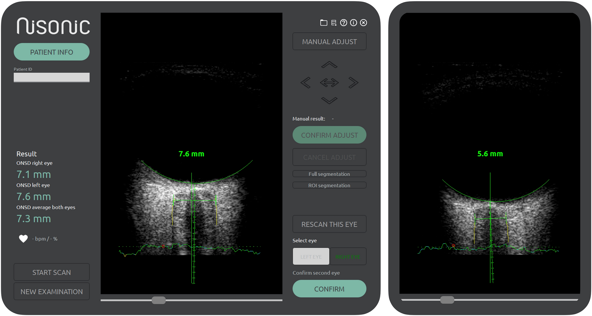
Although several research pieces encourage ultrasonographic ONSD assessment in patients with sepsis, key concerns over its reproducibility, observers’ variations, and measurement accuracy have been raised because all sonographic measurements are operator dependent, and there is a wide range of normal ONSD cutoff levels. Numerous authors previously reported its high level of interobserver and intraobserver reliability. Ballantyne et al. [34] assessed the observer variations in ONSD sonographic measurement of normal adult volunteers with three examiners (one consultant radiologist and two trainees). They reported it to be a reproducible method with low interobserver and intraobserver variability, stressing the importance of standardization of the examination method [34]. Also, Bauerle et al. reported a very high level of intraobserver reliability, reaching 0.92 to 0.97 [35]. Additionally, Wang et al. [36] conducted an investigation with two observers; one observer had 8 years of ultrasonography experience, whereas the other observer was a novice resident who received only 1 week of training in transorbital ultrasonography. They found high interobserver and intraobserver reliability, confirming ONSD sonographic assessment as a highly reproducible and reliable method for ICP evaluation [36]. In concordance with these studies, Kc et al. documented high intraobserver reliability and excellent correlation of interobserver reliability between the two observers [37]. Therefore, ocular ultrasound for ONSD measurement with the standard technique has significant interobserver and intraobserver agreement, confirming its reproducibility and reliability.
In this study, for diagnosing sepsis, the third international definition and appropriate diagnostic criteria, including the SOFA score, were used [2]. The SOFA score is the most common score used and is reported by many studies to have good diagnostic and prognostic predictive value in patients with sepsis [38]. Our research demonstrated significantly high SOFA scores in patients with SAE compared to patients without SAE as well as significantly high APACHE II scores in patients with SAE. Both scores are main indicators of the severity of a patient’s condition, indicating that patients with SAE are more severely ill than patients without SAE [39]. However, their validity to diagnose SAE is still unclear [40, 41]. Patients with higher SOFA and APACHE II scores may be more likely to have SAE, but this may be a biased conclusion because it is worth mentioning that GCS assessment is one of the elements of the SOFA and APACHE II scores, and the diagnosis of SAE is defined by a GCS score < 15.
Patients with SAE tend to have higher ICU-LOS and mortality than those with sepsis alone. Eidelman et al. [42] found that the occurrence of SAE increased hospital mortality from 16% when the GCS score was 15 to 63% when the GCS score was between 3 and 8. Similar conclusions were reported by various studies linking SAE to increased ICU stay and higher short-term mortality [22, 26, 43,44,45]. Our study also demonstrated a significantly extended ICU stay and increased ICU mortality in patients with SAE.
In this study, ONSD measurements of patients with SAE on day 2 showed a positive correlation with both the SOFA score assessments (r = 0.485, P < 0.001) and the ICU-LOS (r = 0.238, P < 0.001), indicating that the wider the ONSD, the more severely ill the patients. In their report, Czempik et al. found no correlation between ONSD and the SOFA score, which could be explained by the different included patients because they investigated only ten patients with septic shock in a mixed ICU, and they used a higher limit of ONSD to diagnose increased ICP (5.7 mm versus 5 mm in our study) [9]. In the present study, wider ONSD was observed in patients who died compared to patients who survived, which is in line with the findings by Yang et al., who demonstrated that patients who died of SAE had slightly wider ONSD than surviving patients [11].
Our results documented that ONSD was helpful in early identification of increased ICP in patients with sepsis and that a wider ONSD reported in patients with SAE could be an early prediction of SAE occurrence, with a cutoff > 5.2 mm. High suspicion of SAE should arise in patients with sepsis with any mental status alterations. ONSD measurement implementation as a screening tool in the diagnostic model could maximize early identification of these patients and stratify the decisions of escalating and individualizing treatment with aggressive measures to optimize cerebral perfusion pressure. Therefore, future studies with efforts to include populations from different regions and different ethnic groups and using standardized methods to assess and implement ONSD measurement in the sepsis management protocol are highly required.
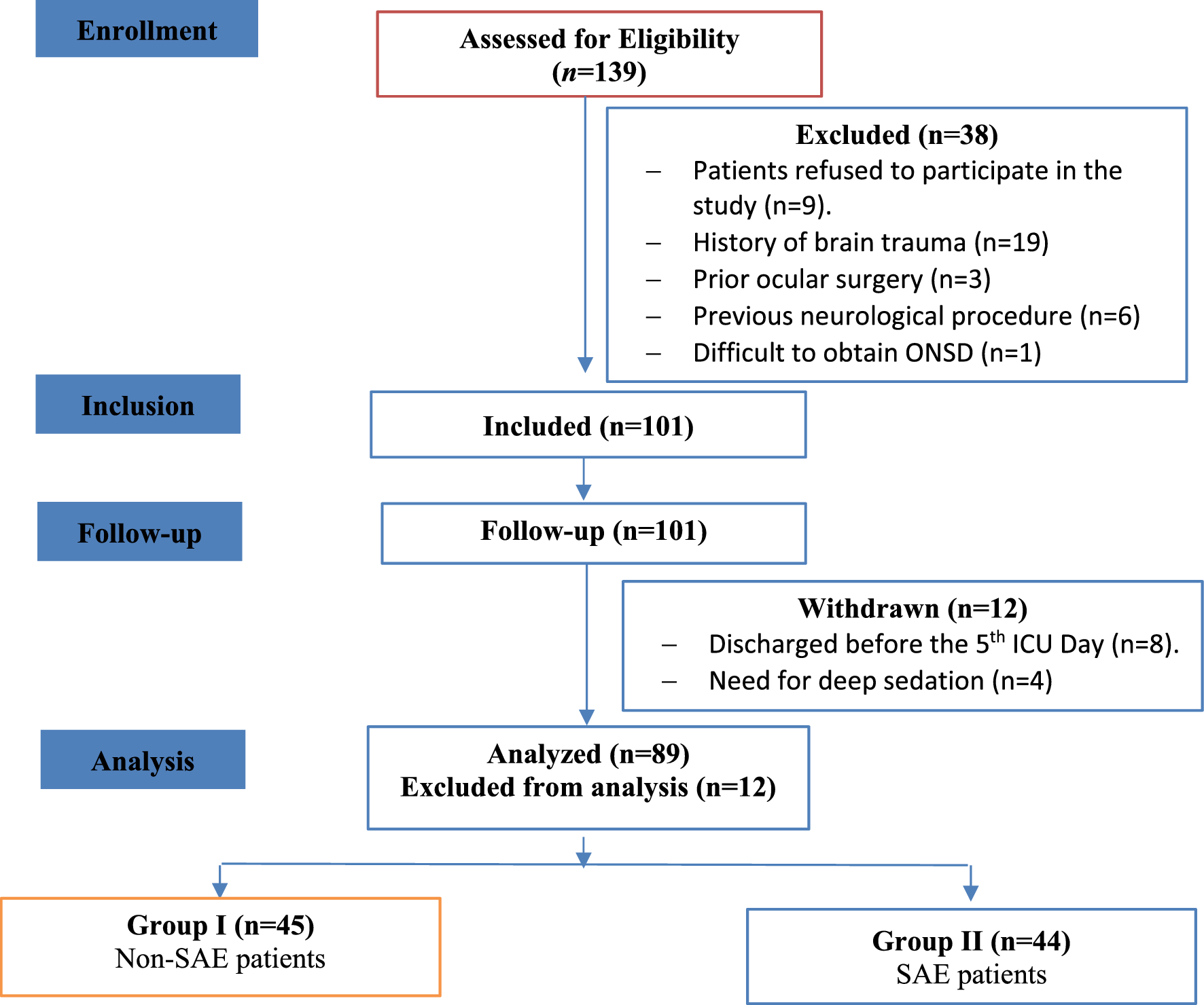
However, this study has some limitations. Firstly, studying SAE is challenging because there are no specific diagnostic criteria, and it remains a diagnosis of exclusion. Although we used a GCS score < 15 and a positive CAM-ICU result, which are relatively objective methods, the diagnosis by ICU physicians based mainly on clinical symptoms of SAE may be somewhat subjective. Hence, missed diagnoses or misdiagnoses might occur. Second, with more than one examiner, errors may be encountered in results interpretation. Moreover, reproducibility and minimizing observers’ variation are crucial concerns in using ONSD sonographic assessment for repeated monitoring. We established a unified standard ONSD measurement method for all our patients to be used by an experienced operator (our two examiners had the same level of experience on ocular ultrasound and used the same examination technique). Standardizing the examination method could help to maintain consistency and reduce observers’ variation. Perfectly, such technical factors standardization may help perform future well-designed clinical studies to evaluate ultrasonographic ONSD benefits.


Comments (0)