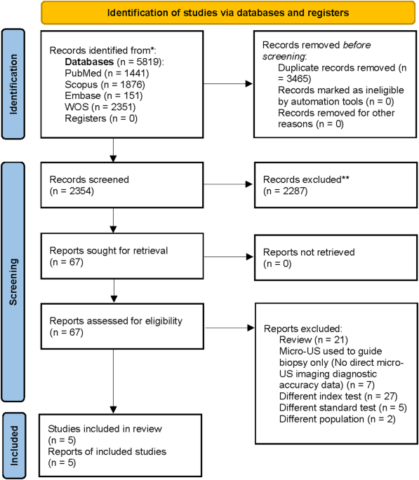
This systematic review and meta-analysis evaluated 29-MHz micro-ultrasound (micro-US, ExactVu) as a stand-alone index test for classifying clinically significant prostate cancer (csPCa) across five prospective studies. The random-effects model on logit-transformed sensitivity and specificity showed high pooled sensitivity and low pooled specificity, sensitivity 0.84 (95% CI 0.65–0.94) and specificity 0.41 (95% CI 0.25–0.59), with an HSROC consistent with moderate discrimination. Secondary metrics were concordant (PLR 1.45, 95% CI 1.17–1.80; NLR 0.37, 95% CI 0.23–0.61; DOR 3.95, 95% CI 2.48–6.30). On a 25% pre-test probability, Fagan analysis indicated modest probability shifts (~ 33% after a positive micro-US; ~11% after a negative), supporting a triage/rule-out role rather than definitive rule-in.
Heterogeneity was substantial across metrics, and a strong negative correlation between logit-sensitivity and logit-specificity indicated threshold effects as a principal driver of variability. Exploratory meta-regression did not identify robust moderators of sensitivity; spectrum-related covariates were associated with lower specificity but given the small number of studies these findings are hypothesis-generating. Model diagnostics supported the adequacy of the bivariate specification, and Deeks’ test showed no evidence of small-study effects. Overall, micro-US demonstrates high sensitivity and low specificity with modest clinical impact on post-test probability, best positioned as a complementary/triage tool, particularly when mpMRI is unavailable, contraindicated, or delayed, rather than a stand-alone confirmatory test.
The pooled sensitivity of micro-US in our analysis was 84%, indicating that micro-US effectively identifies a substantial proportion of patients with PCa. This finding aligns with the results reported in several of the included studies. For instance, Klotz et al.[9] reported a sensitivity of 94% in a large multicenter registry study including 1,040 patients, while Avolio et al. [15] found a sensitivity of 85% in a prospective cohort of 1,423 men undergoing biopsy. Conversely, Pavlovich et al. [16] reported a lower sensitivity of 60%, which may be explained by the inclusion of early operator learning phases and a more heterogeneous patient population. Our pooled sensitivity is also consistent with previous reviews and meta-analyses. Alghamdi et al. [17], in a meta-analysis comparing multiple US modalities, reported pooled sensitivities for US ranging from 74% to 94%, although their analysis combined micro-US with other US techniques and did not assess micro-US in isolation. Similarly, Ditonno et al. [18] reported favorable sensitivity for micro-US but pooled results across heterogeneous imaging approaches, limiting direct comparisons. The high sensitivity of micro-US can be attributed to its high-frequency (29 MHz) real-time imaging, which enables improved visualization of prostate tissue microstructures. This suggests that micro-US may be an effective tool for early lesion detection, potentially reducing missed clinically significant cancers, especially in settings where MRI is unavailable or contraindicated.
The pooled specificity of micro-US was 41% with study-level estimates spanning (25–59%). This represents low specificity, close to chance performance—and argues against using micro-US as a stand-alone rule-in test. In contrast, Lughezzani et al. reported a specificity of 60.6%, and Klotz et al. observed a specificity of 22%, reflecting real-world variability [9, 19]. Previous meta-analyses have also highlighted the challenge of achieving high specificity with US-based modalities. In Alghamdi et al.‘s review, specificity values for US varied widely and were generally lower than those reported for MRI, emphasizing the inherent limitations of US-based imaging for tissue characterization [17]. The low specificity implies more false positives and potential unnecessary biopsies; while standardized scoring (PRI-MUS) and reader experience may improve performance, stricter thresholds that raise specificity generally reduce sensitivity, reinforcing a triage/rule-out rather than confirmatory role for micro-US.
The pooled PLR of 1.45 and NLR of 0.37 suggest that micro-US has limited value for confirming PCa but may offer moderate utility in ruling it out. These values indicate that patients with PCa are approximately 1.4 times more likely to have a positive micro-US result, while a negative result moderately decreases the probability of disease. The Fagan nomogram, using a pre-test probability of 25%, showed that a positive micro-US result increases the post-test probability to 33%, whereas a negative result reduces it to 11%. Accordingly, the likelihood ratio scatter plot places micro-US in the rule-out-leaning zone, consistent with a “rule-out” function rather than confirmatory use. The pooled DOR of 3.95 reflects modest overall discriminative ability. From a clinical standpoint, these findings indicate that micro-US, while sensitive, lacks the specificity needed to reliably confirm disease and is best positioned as an exclusion tool, particularly in settings where mpMRI is unavailable, contraindicated, or delayed. However, the substantial heterogeneity across studies (I² >80% for sensitivity, specificity and DOR) underscores variability in test performance and highlights the need for standardized operator training, consistent scoring systems (e.g., PRI-MUS) [16, 20], and consideration of disease prevalence when applying micro-US in routine clinical decision-making.
Substantial heterogeneity in diagnostic accuracy was observed and should temper interpretation of the pooled estimates. From a clinical perspective, such variability is unsurprising: imaging performance depends on patient spectrum, reader experience, and local protocols. Our analyses demonstrated a clear threshold effect, with a trade-off between sensitivity and specificity that likely reflects differences in positivity criteria (e.g., PRI-MUS cut-offs) and reader behavior. In subgroup summaries, sensitivity was broadly stable across strata, whereas specificity varied with clinical spectrum: cohorts enriched for higher pre-test risk (such as prior-negative biopsy populations or screening-type case mixes) tended to show lower and more variable specificity, and single-study strata often combined very high sensitivity with very low specificity, consistent with threshold/spectrum phenomena and warranting cautious interpretation. In meta-regression, no study characteristic consistently shifted sensitivity, while indicators of enriched spectrum were associated with lower specificity; by contrast, factors such as sample size, center type, biopsy approach or guidance, reference standard, and geography did not explain residual variability. Taking them together, these findings suggest that heterogeneity is driven predominantly by thresholding and spectrum rather than by any single procedural detail. Standardizing PRI-MUS thresholds, reinforcing reader training, and clearly defining target populations may improve calibration; clinically, micro-US is most defensible as a triage/rule-out tool in pathways where rapid, accessible assessment is needed or mpMRI access is limited, with explicit acknowledgment of the sensitivity–specificity trade-off.
Comparison with mpmri: diagnostic accuracy and feasibility
Our pooled micro-US performance (sensitivity 0.84; specificity 0.41) should be interpreted in the context of contemporary mpMRI. In the paired-design PROMIS study using template mapping as the reference, mpMRI achieved high sensitivity for clinically significant cancer (≈ 93%) with lowest specificity (≈ 41%), broadly like the specificity we observed for micro-US but with higher sensitivity overall [21]. In larger evidence syntheses, a 2019 meta-analysis of 29 prospective mpMRI studies (8,503 men) reported pooled sensitivity of 0.87 and specificity of 0.68 for suspected prostate cancer, indicating that mpMRI typically operates on a more favorable sensitivity–specificity frontier than micro-US in our analysis [22]. Current guidelines therefore endorse an MRI-first pathway before biopsy in biopsy-naïve men, reflecting both diagnostic performance and the ability of mpMRI to guide targeted sampling.
Feasibility considerations, however, may favor complementary roles. mpMRI requires specialized scanners, protocols, and radiology expertise, with longer scheduling and higher costs, whereas micro-US is bedside, real-time, contrast-free, and can be integrated into the same-day diagnostic visit. Against this, our meta-analysis shows that micro-US specificity is low, implying more false positives if used as a stand-alone rule-in test. A pragmatic interpretation is that micro-US could serve as a rapid triage/rule-out adjunct where mpMRI is unavailable, contraindicated, or delayed, or as an access-expanding gatekeeper to MRI in resource-constrained settings. Determining the optimal sequencing or combination (e.g., micro-US–guided targeting with or without MRI) will require prospective head-to-head or combined-strategy studies powered for clinically significant endpoints [23].
Cost and resource implications
Formal, peer-reviewed economic evaluations directly comparing stand-alone micro-ultrasound with mpMRI are currently scarce. By contrast, multiple analyses suggest that mpMRI-first pathways are cost-effective versus systematic TRUS biopsy alone because they reduce unnecessary biopsies and overdiagnosis. For example, a Singapore health-system model found pre-biopsy MRI strategies to be cost-effective relative to TRUS-only pathways across plausible willingness-to-pay thresholds, primarily by improving detection of clinically significant cancer and avoiding low-yield biopsies [24]. In settings where scanner capacity is constrained, abbreviated/shorter MRI protocols have also been reported as highly cost-effective compared with full mpMRI while maintaining diagnostic performance, further supporting MRI-first pathways from an economic standpoint. Representative unit-cost data used in screening and diagnostic models (e.g., UK analyses drawing on NICE resource estimates) place mpMRI as a relatively high-cost diagnostic compared with ultrasound-based approaches, reinforcing that per-examination costs and infrastructural needs are materially greater for MRI [25].
Micro-ultrasound, as an ultrasound-based, contrast-free, point-of-care modality, is inherently less resource-intensive in terms of equipment, scheduling, and on-site logistics; early prospective trials comparing micro-US–guided pathways with MRI-based strategies suggest potential for wider accessibility and lower direct costs, though these signals come largely from feasibility and non-economic reports to date [26]. Taken together, the current evidence supports mpMRI as cost-effective versus TRUS-only pathways, while the cost-effectiveness of micro-ultrasound—either as a stand-alone alternative or as a triage gatekeeper to mpMRI—remains an open question. Prospective economic evaluations that quantify quality-adjusted life-years, downstream biopsy utilization, and incremental cost-effectiveness ratios for micro-US–first or combined micro-US/mpMRI strategies are needed to define the most efficient deployment across health-system contexts.
Strengths and limitations
This work is, to our knowledge, among the first pooled diagnostic-test-accuracy syntheses focused exclusively on 29-MHz micro-ultrasound as a stand-alone index test for classifying clinically significant prostate cancer (csPCa) in prospective cohorts. We limited inclusion to studies using histopathology obtained in the same diagnostic episode and a standardized platform (ExactVu), which improves comparability while recognizing remaining spectrum and threshold differences. We applied a random-effects pooling of logit-transformed sensitivity and specificity with an HSROC representation and pre-specified thresholds with pre-specified thresholds (primary PRI-MUS ≥ 3, secondary ≥ 4), presented HSROC curves, and conducted comprehensive diagnostics (leave-one-out influence, goodness-of-fit, and residual checks). Subgroup summaries and meta-regression were used to explore heterogeneity, and sensitivity analyses (e.g., excluding the spectrum-selected cohort and the randomized guidance trial) assessed robustness. Finally, we quantified clinical impact with likelihood ratios, DOR, and Fagan nomograms, providing decision-relevant interpretation for real-world pathways where rapid triage or limited mpMRI access is a concern.
Several limitations merit consideration. The evidence base remains small (five prospective studies), which limits precision and renders subgroup and meta-regression results exploratory rather than confirmatory. Between-study heterogeneity was substantial across all metrics and appears driven chiefly by thresholding (differences in PRI-MUS cut-offs and reader behavior) and clinical spectrum (e.g., enrichment by prior-negative biopsy or MRI-negative pathways), with one included randomized guidance trial contributing design diversity; although leave-one-out checks suggested no single study dominated the results, pooled estimates should be interpreted cautiously. Generalizability may be constrained because most cohorts were from experienced, often academic centers using a single platform (ExactVu), and performance can vary with operator training and learning curve. Despite using histopathology as the reference standard, variations in biopsy approach (transrectal vs. transperineal), sampling strategy (systematic, targeted, or combined), guidance modality, and csPCa definitions (e.g., ISUP ≥ 2 vs. alternatives) may introduce verification and classification differences that our study-level moderators could not fully capture. Although Deeks’ test did not indicate small-study effects, the low power inherent to few studies means selective reporting or publication bias cannot be excluded. Future research should prioritize multicenter, prospective DTA designs with pre-specified PRI-MUS thresholds, explicit blinding, and uniform verification irrespective of test result; include community settings; perform head-to-head and combined-strategy evaluations with mpMRI; and report decision-curve and cost-effectiveness outcomes to clarify the most efficient role for micro-US in routine pathways.
Conclusion
Micro-US shows high sensitivity but low specificity for classifying clinically significant prostate cancer, resulting in modest overall discrimination and only modest shifts in post-test probability. In practice, it is best used as a triage/rule-out adjunct, especially when mpMRI is unavailable, contraindicated, delayed, or when same-day assessment is needed, rather than as a stand-alone rule-in test; its performance is sensitive to threshold and clinical spectrum, reinforcing the need for standardized PRI-MUS application and reader training.





Comments (0)