Wong TY, Sabanayagam C. Strategies to tackle the global burden of diabetic retinopathy: from epidemiology to artificial intelligence. Ophthalmologica. 2020;243:9–20.
Article
CAS
PubMed
Google Scholar
Ansari P, Tabasumma N, Snigdha NN, Siam NH, Panduru RVNRS, et al. Diabetic retinopathy: an overview on mechanisms, pathophysiology and pharmacotherapy. Diabetology. 2022;3:159–75.
Article
Google Scholar
Forrester JV, Kuffova L, Delibegovic M. The role of inflammation in diabetic retinopathy. Front Immunol. 2020;11:1–22.
Borrelli E, Sacconi R, Brambati M, Bandello F, Querques G. In vivo rotational three-dimensional OCTA analysis of microaneurysms in the human diabetic retina. Sci Rep. 2019;9:16789. https://doi.org/10.1038/s41598-019-53357-1.
Akram MU, Khalid S, Khan SA. Identification and classification of microaneurysms for early detection of diabetic retinopathy. Pattern Recognit. 2013;46:107–16.
Article
Google Scholar
Horii T, Murakami T, Nishijima K, Sakamoto A, Ota M, Yoshimura N. Optical coherence tomographic characteristics of microaneurysms in diabetic retinopathy. Am J Ophthalmol. 2010;150:840–8.
Querques G, Borrelli E, Battista M, Sacconi R, Bandello F Optical coherence tomography angiography in diabetes: focus on microaneurysms. Eye. 2020;35 https://pubmed.ncbi.nlm.nih.gov/32887935/.
Borrelli E, Battista M, Sacconi R, Querques G, Bandello F. Optical coherence tomography angiography in diabetes. Asia Pac J Ophthalmol. 2021;10.
Borrelli E, Sacconi R, Parravano M, Costanzo E, Querques L, Battista M, et al. OCTA assessment of the diabetic macula: a comparison study among different algorithms. Retina. 2021.
Kaizu Y, Nakao S, Wada I, Arima M, Yamaguchi M, Ishikawa K, et al. Microaneurysm imaging using multiple En face OCT angiography image averaging: morphology and visualization. Ophthalmol Retina. 2020.
Karst SG, Salas M, Hafner J, Scholda C, Vogl W-D, Drexler W, et al. Three-dimensional analysis of retinal microaneurysms with adaptive optics optical coherence tomography. Retina. 2019.
Parravano M, De Geronimo D, Scarinci F, Querques L, Virgili G, Simonett JM, et al. Diabetic microaneurysms internal reflectivity on spectral-domain optical coherence tomography and optical coherence tomography angiography detection. Am J Ophthalmol.2017;179:90–6. https://linkinghub.elsevier.com/retrieve/pii/S0002939417301903.
Parravano M, De Geronimo D, Scarinci F, Virgili G, Querques L, Varano M, et al. Progression of diabetic microaneurysms according to the internal reflectivity on structural optical coherence tomography and visibility on optical coherence tomography angiography. Am J Ophthalmol. 2019;198:8–16. http://www.ncbi.nlm.nih.gov/pubmed/30308201.
Arrigo A, Teussink M, Aragona E, Bandello F, Battaglia Parodi M. MultiColor imaging to detect different subtypes of retinal microaneurysms in diabetic retinopathy. Eye. 2021;35:277–81.
Article
CAS
PubMed
Google Scholar
Parravano M, De Geronimo D, Scarinci F, Querques L, Virgili G, Simonett JM, et al. Diabetic microaneurysms internal reflectivity on spectral-domain optical coherence tomography and optical coherence tomography angiography detection. Am J Ophthalmol. 2017;179:90–6.
Article
PubMed
Google Scholar
Sun Z, Yang D, Tang Z, Ng DS, Cheung CY. Optical coherence tomography angiography in diabetic retinopathy: an updated review. Eye. 2021;35:149–61.
Article
PubMed
Google Scholar
Borrelli E, Grosso D, Barresi C, Lari G, Sacconi R, Senni C, et al. Long-term visual outcomes and morphologic biomarkers of vision loss in eyes with diabetic macular edema treated with anti-VEGF Therapy. Am J Ophthalmol. 2021. https://pubmed.ncbi.nlm.nih.gov/34509431/.
Vujosevic S, Aldington SJ, Silva P, Hernández C, Scanlon P, Peto T, et al. Screening for diabetic retinopathy: new perspectives and challenges. Lancet Diabetes Endocrinol. 2020;8:337–47.
Article
PubMed
Google Scholar
Oakley JD, Verdooner S, Russakoff DB, Brucker AJ, Seaman J, Sahni J, et al. Quantitative assessment of automated optical coherence tomography image analysis using a home-based device for self-monitoring neovascular age-related macular degeneration. Retina. 2022.
Ronneberger O, Fischer P, Brox T. U-Net: Convolutional networks for biomedical image segmentation BT - medical image computing and computer-assisted intervention – MICCAI 2015. In: Navab N, Hornegger J, Wells WM, Frangi AF, editors. Cham: Springer International Publishing; 2015. p. 234–41.
Oakley JD, Sodhi SK, Russakoff DB, Choudhry N. Automated Deep Learning-based Multi-class Fluid Segmentation in Swept-Source Optical Coherence Tomography Images. 2020. https://doi.org/10.1101/2020.09.01.278259.
Borrelli E, Oakley JD, Iaccarino G, Russakoff DB, Battista M, Grosso D, et al. Deep-learning based automated quantification of critical optical coherence tomography features in neovascular age-related macular degeneration. Eye. 2023.
Ricardi F, Oakley J, Russakoff D, Boscia G, Caselgrandi P, Gelormini F, et al. Validation of a deep learning model for automatic detection and quantification of five OCT critical retinal features associated with neovascular age-related macular degeneration. Br J Ophthalmol. 2024: bjo-2023-324647.
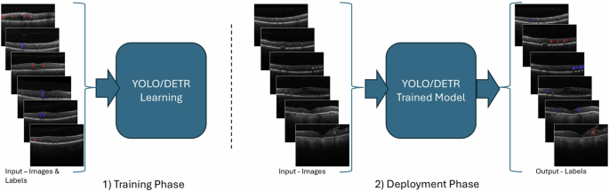
Redmon J, Divvala S, Girshick R, Farhadi A. You only look once: unified, real-time object detection. http://pjreddie.com/yolo/.
Bochkovskiy A, Wang C-Y, Liao H-YM. YOLOv4: optimal speed and accuracy of object detection. 2020. http://arxiv.org/abs/2004.10934.
Zhu X, Su W, Lu L, Li B, Wang X, Dai J. Deformable DETR: deformable transformers for end-to-end object detection. 2020. http://arxiv.org/abs/2010.04159.
YOUDEN WJ. Index for rating diagnostic tests. Cancer. 1950;3:32–5.
Article
CAS
PubMed
Google Scholar
Almasi R, Vafaei A, Kazeminasab E, Rabbani H. Automatic detection of microaneurysms in optical coherence tomography images of retina using convolutional neural networks and transfer learning. Sci Rep. 2022;12:13975.
Article
CAS
PubMed
PubMed Central
Google Scholar
Anon. https://github.com/Shenggan/BCCD_Dataset.
Gulshan V, Peng L, Coram M, Stumpe MC, Wu D, Narayanaswamy A, et al. Development and validation of a deep learning algorithm for detection of diabetic retinopathy in retinal fundus photographs. JAMA. 2016;316:2402–10. https://jamanetwork.com/journals/jama/fullarticle/2588763.
Abràmoff MD, Lou Y, Erginay A, Clarida W, Amelon R, Folk JC, et al. Improved automated detection of diabetic retinopathy on a publicly available dataset through integration of deep learning. Invest Ophthalmol Vis Sci. 2016;57:5200–6.
Article
PubMed
Google Scholar
Pratt H, Coenen F, Broadbent DM, Harding SP, Zheng Y. Convolutional neural networks for diabetic retinopathy. In: Procedia computer science. Elsevier B.V. 2016;90:200–5.
Abràmoff MD, Lavin PT, Birch M, Shah N, Folk JC. Pivotal trial of an autonomous AI-based diagnostic system for detection of diabetic retinopathy in primary care offices. NPJ Digit Med. 2018;1.
Parravano M, De Geronimo D, Scarinci F, Virgili G, Querques L, Varano M, et al. Progression of diabetic microaneurysms according to the internal reflectivity on structural optical coherence tomography and visibility on optical coherence tomography angiography. Am J Ophthalmol. 2019;198:8–16.
Article
PubMed
Google Scholar
Zhang L, Van Dijk EHC, Borrelli E, Fragiotta S, Breazzano MP. OCT and OCT angiography update: clinical application to age-related macular degeneration, central serous chorioretinopathy, macular telangiectasia, and diabetic retinopathy. Diagnostics. 2023;13:232.
Article
PubMed
PubMed Central
Google Scholar


Comments (0)