
Remember me


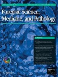

From the 144 articles obtained, 14 were included for analysis and data extraction (Fig. 1). These were experimental studies, published in English, and presented a low risk of bias analysis (Supplementary Table 1). All articles assessed the changes in microbial communities throughout the cadaveric decomposition process and their use for estimating PMI (Table 1). Twelve articles explored the bacterial community [7, 12,13,14, 20,21,22,23,24,25,26,27]. From those, the bacterial community was evaluated by 16S rRNA gene amplicon high-throughput sequencing in 11 articles [7, 12,13,14, 20,21,22,23,24,25,26,27]; by qPCR targeting Bacteroides, Lactobacillus, and Bifidobacterium in one paper [23]; by 16S rRNA gene high-throughput sequencing combined with metagenomic and metatranscriptomic sequencing and culture in selective and rich media in one paper [27]; and by 16S rRNA and 18S rRNA amplicon high-throughput sequencing to explore also the eukaryotic communities in another [26]. One paper explored the eukaryotic fungi ecosystem through culture in selective media and photography to macroscopically monitor the area of fungal coverage [28], and another paper evaluated the microbial neoformation of volatiles using headspace gas chromatography with flame ionization identification method (HS-GC-FID) [29]. Moreover, different studies focused on the microbial ecology of other anatomic areas: head external orifices (hard palate, mouth, nose, ears, and eyes) [13, 21, 24, 25, 27], gut [12, 23], internal organs and blood [14, 29], skin [28], and bones [22, 26]. Also, the interactions between corpse and soil microbiomes were accessed [7, 20, 22, 26].
Fig. 1
Systematic review flowchart leading to selection of articles
Table 1 Summarisation of the information obtained from the articles under analysisPMI was measured in hours, days, months, or years or as accumulated degree days (ADD). ADD is the cumulative total of daily average temperatures. Linking the decomposition stages or insect development to ADD allows temperature variations to be considered when estimating PMI [31]. Cumulative degree hours (CDH), a refinement of the ADD calculation process for cadavers decomposing over a short period, represent an average value for each 12-h interval. Similarly to ADD calculation, temperatures below 0 °C are counted as a zero, and negative values are not used [32, 33]. The Total Body Score method (TBS), a scale that distinguishes decomposition different stages, allowing to assign points to specific categories and eventually to score overall decomposition [34, 35], was also used [29].
For clarity, the description of the individual studies will be organized by the anatomical areas, when possible. For detailed and organized data, please refer to Table 1. Adserias-Garriga et al. [21] took oral swab samples from three cadavers at different putrefaction stages; all showed similar successional changes, despite having other oral conditions. Bacillota (previously named Firmicutes) and Actinomycetota (previously called Actinobacteria) were the most predominant phyla from day 1 to 5, coinciding with the fresh stage. Their relative abundances decreased from day 1 to day 5–6, whereas Tenericutes (gastro-intestinal microbiota) appeared at the bloat stage (days 5 and 7). The oral microbiome was characterized: i) in the fresh stage by indigenous oral commensals; ii) in the bloat stage by both oral (Peptostreptococcaceae, Bacteroidaceae) and gut (Enterococcaceae, and Clostridiales) microbiota; iii) in advance decay, mainly by soil microbiota; and iv) in dry remains, by bacilli and clostridia. The authors suggest oxygen availability is a determinant factor for bacterial ecological changes.
Ashe et al. [27] sampled three cadavers’ hard palate, and samples were analyzed by combining 16S rRNA sequencing with whole metagenomic (MetaG) and metatranscriptomic (MetaT) sequencing and cultured in selective and rich media. The analysis by different sequencing techniques allowed a more complete view of the microorganisms present at the various decomposition. Concerning the 16S taxonomic distributions, 25 to 70% were Bacillota in the fresh decomposition phase (ADD values up to 50); Pseudomonadota (previously named Proteobacteria) were found in most samples, and Actinomycetota were more numerous in 2 out of 7 samples; in one sample, Actinomycetota consisted of 75% due to the genus Rothia. Middle to later decomposition stages (> ADD 168) reveal that Bacillota dominated (> 75% sequences) or co-dominated (~ 50% sequences) in 9 of 13 samples; Pseudomonadota dominated or co-dominated in 5 of 13 samples, and in one sample (291 ADD), Actinomycetota registered more than 50% of sequences. Pseudomonas spp. was most common in later samples. Regarding MetaG Taxonomic distributions, Streptophyta (from Plantae kingdom) were common (11 of 17 samples), Bacillota were found in earlier samples, and Pseudomonadota were higher in later samples. On the taxonomic distributions of MetaT, bacterial data showed that Bacillota were common for the early and middle samples, Pseudomonadota were generally lower (except for the later stages samples); Actinomycetota were found in lower numbers in both early and later samples. Cultures included 46 bacterial unique species from 69 isolates. Species from the Pseudomonadota accounted for the isolates’ largest proportion (43.5%). Bacillota (32.6%), Actinomycetota (19.6%), and Bacteroidota (previously named Bacteroidetes) (4.3%) accounted for the rest. In total, 4 phyla were identified by culture, 7 by 16S sequencing, 66 by MetaG sequencing, and 51 by MetaT sequencing, more than half bacterial. Bacillota, Pseudomonadota, and Actinomycetota were the most abundant. Still, their distributions did not distinguish samples based on decomposition stage or time or by donor. However, Bacillota were more common in the early and Pseudomonadota in the later stages. Better resolution was observed at the genus level, where a distinction could often be made between the community from fresh (ADD < 50) vs. later (ADD > 168) sampling times. Taxa from the standard living human oral microbiome (e.g., Lactobacillus, Streptococcus, Rothia, and Candida) were replaced as decomposition progressed by Lysinibacillus, Vagococcus, Ignatzschineria, and Yarrowia. Still, a relationship between PMI and the bacterial community present during sampling was not established since only differentiation between fresh and late states was considered.
Another study [25] aimed to evaluate the machine learning methods’ performance on microbial community analysis obtained from 188 cases and postmortem samples from different anatomical areas (ears, eyes, nose, mouth, and rectum). Machine learning methods are tools that implement neural network and random forest models to perform regression and feature selection tasks on microbiome data; these methods allow the assessment of large multi-dimensional datasets that otherwise would be difficult to analyze and interpret. In the study mentioned above, for PMI prediction (i.e., their abilities to predict PMI), the highest accuracy (77.5%) was achieved when all anatomic areas were used. The most relevant microbial taxa identified were Veillonella dispar and Proteus sp. (PMI > 73 h). In comparison, Moraxellaceae had a higher count in cases with 49–72 h estimated PMI, and Streptococcus sp. within 48 h PMI. Thus, microbial signatures change between the time frames less than 2 days postmortem and more than 2 days postmortem, with an increasing number of unique taxa associated with communities in the first 2 days. As decomposition progresses, the postmortem richness (the total number of species in a community) and diversity (the number of species and their abundance in a community) of the microbial community decrease significantly (PMI ≥ 48 h).
Johnson et al. [24] also used a machine-learning approach, with 114 microbial samples collected from 21 cadavers’ ears and nasal cavities. The best methods and PMI indicators were evaluated to establish an algorithm to predict PMI from microbial samples, using three computational methods: F-value (feature selection considers the coefficient resulting from fitting a single feature with the target using a linear model), a tree-based approach (ranks features on their tendency to occupy essential positions in decision trees built on the same dataset), and mutual information (scores each taxon on the amount of information it has in common with the dependent variable). The results showed that the entire data set analysis works best, rather than any specific taxon or even small group of taxa; some taxa were identified as powerful indicators of PMI: phyla Actinomycetota and Armatimonadota and the classes Thermoleophilia and Erysipelotrichi were marked as top results by all three methods; the orders Myxococcales and Erysipelotrichales, the families Staphylococcaceae, Planococcaceae, and Enterococcaceae, and genera Staphylococcus and Vagococcus were identified as essential features by two of three methods. A correlation between microbial diversity and ADD was proven; the ear microbiome diversity was negatively correlated with ADD, and the nasal microbiome was positively correlated but less pronounced. Additionally, analyzing taxon at multiple levels simultaneously and combining samples from different body sites improved the model’s accuracy.
In the Hauther et al. study [23], Bacteroides, Lactobacillus, and Bifidobacterium were quantified in the gut by qPCR using targeted primers in 12 cadavers (including 6 controls, sampled only once). For 20 days, corresponding to 600 CHD, Bacteroides and Lactobacillus relative abundances declined exponentially with increasing PMI. Contrarily, PMI did not significantly alter Bifidobacterium abundance. Repeated sampling affected Bifidobacterium abundance, likely due to oxygen introduction but not Bacteroides and Lactobacillus populations. This study shows that the gut Bacteroides and Lactobacillus populations may be used as a PMI indicator at these intervals, as both populations decline with PMI.
The DeBruyn and Hauther study [12] also investigated the postmortem changes in the gut microbiome of 4 cadavers for 30 days after death. The bacterial communities’ taxon richness increased with time while the diversity decreased. An “early” and “late” microbial structure was defined, with changes occurring at the bloat stage (4 to 7 days). In the “early” microbial communities, the diversity was high, with the predominant phyla being those characteristics of the human gut microbiome: Bacteroidota and Bacillota; the “late” microbial communities had a higher richness but had lower diversity since the relative abundance of Bacteroidota declines. Although Bacillota still dominated, these communities were significantly enriched with microorganisms belonging to Clostridiales order and the fly-associated Gamma-Proteobacteria. Bacteroides and Parabacteroides declined over time and were significantly inversely correlated to PMI, and Clostridium was a PMI strong positive predictor.
In the Lutz et al. study [14], postmortem microbial DNA was extracted from several organs (brain, heart, liver, spleen, prostate, and uterus) of 40 Italian cadavers to investigate variation and microbial associations among different body organs in human cadavers to predict PMI. The different organs’ bacterial communities’ analysis showed significant differences in the relative abundances of multiple taxa, as the non-reproductive organs were dominated by bacterial orders MLE1-12, Saprospirales and Burkholderiales. In contrast, reproductive organs were dominated by Clostridiales and Lactobacillalaes and showed a marked decrease in relative abundance of MLE1-12. Several significant relationships were identified regarding the association between PMI and bacterial relative abundances. Within the heart, taxa belonging to the order Burkholderiales exhibited a significant increase in relative abundance with increasing PMI. In all organs, except for the uterus, taxa belonging to the order Clostridiales demonstrated an increase in relative abundance with increasing PMI (only significant for the brain, liver, and spleen). In the brain, heart, liver, and spleen, taxa of the order MLE1-12 showed a slight decrease in relative abundance with increasing PMI but not significant.
Javan and colleagues [13] analyzed the 20 predominant bacterial genera relative abundances in 66 samples of different human body organs (brain, mouth cavity, heart, liver, and spleen) and blood collected from 27 human corpses, with a PMI between 3.5 and 240 h. The thanatomicrobiome signatures showed time-, organ-, and sex-dependent changes useful for PMI estimation. Family and genus-level analyses explained approximately 21% of the variance in models correlating PMI, while species-level study explained 65%. Several genera, such as Clostridium and Prevotella, could predict different decomposition periods. The phylum Bacillota was identified as a possible biomarker in the thanatomicrobial communities from other body locations. Although the phylum was not a strong PMI predictor, Bacillota genera such as Clostridium, Bacillus, Peptoniphilus, Blautia, and Lactobacillus exhibit temporal changes. Bacterial genera were similar among different organs within each sex but dissimilar between females and males, except in the oral cavity. Females had a higher relative abundance of Pseudomonas and Clostridiales, while males had Clostridium, Clostridiales, Streptococcus, and Rothia.
Ceciliason et al. [29] analyzed blood collected from 412 corpses to assess the presence of ethanol, N-propanol, 1-butanol, and acetaldehyde. The volatiles were analyzed by HS-GC-FID. The decomposition degree was evaluated in three anatomical regions based on external visual signs. The obtained dataset was divided into non-decomposition and external decomposition cases, and PMI ranged from 0 to 6 days in the first group and 0 to 106 days in the second. The most common microbial-derived volatiles found were acetaldehyde (83%), followed by ethanol (37%), N-propanol (21%), and 1-butanol (4%). The results indicated no or weak linear relationship between PMI and detected volatiles, probably because this study accessed an indirect measure of the microbial community.
Fungal colonies were identified on the cadaver surface (skin), and their macromorphological variation was used to assess their potential as a PMI marker [28]. Photographs to monitor the fungal coverage area were made, and samples were collected for culture and identification of fungi. Initially, fungal colonies appeared on the face, right arm, and both ankles; during the following weeks, the colonies spread to the arms, chest, abdomen, autopsy suture, groin, and legs. In the more colonized areas, the corpse tissues appear to have a higher level of dehydration compared with the non-colonized areas. Also, a quite homogeneous and gradual growth on the face was observed and accompanied by a chromatic variation of each colonized area in relation to the time development. In another case, fungal growth was limited to the oral cavity, and the colonies observed were Penicillium expansum and Cladosporium cladosporioides, with colonization beginning on the palate 2 days after death. The colonization pattern diversity is caused by the interaction of the initial inoculum, initial conditions of the corpse, and the environmental parameters in the perimortem period; therefore, the corpse mycobiota characterization may also reveal possible changes in the corpse’s location.
Damann’s study [22] aimed to evaluate the bacterial communities’ potential as a method for estimating PMI for long periods, assess the differences in the microbiome between partially skeletonized remains (PMI = 27–284 d (days)), skeletonized samples (PMI = 292–369 d), and dry remains (PMI = 554–1692 d). In parallel, soil samples were collected for comparison to the ribs microbiome. Partially skeletonized remain samples, followed by the fully skeletonized, presented the lowest taxonomic diversity level. Pseudomonadota was the most abundant in all sample groups. Alpha-Proteobacteria increased relative abundance for each successive bone decay stage, while Gamma-Proteobacteria decreased. After the Pseudomonadota, members of the Bacillota and Bacteroidota were the most abundant, mostly present in the gut microbiota. Actinomycetota, prevalent in gut and soil microbiomes, and Acidobacteriota, present exclusively in the environment, were more prevalent in dry than partially/fully skeletonized remains. The partially skeletonized remains (PMI:27–284 days) presented the highest proportion of Bacillota; the fully skeletonized remains (PMI:292–369 days) showed the highest relative abundance of Bacteroidota; and the dry remains (PMI:554–1692 days) hosted the most significant proportion of Actinomycetota. The interstage taxonomic succession from decaying bone suggested an underlying continuous transition in community composition, partially skeletonized remains maintained a presence of bacteria associated with the human gut, and the dry skeletal remains bacterial composition kept a community profile like soil communities. This work also showed that community membership (unweighted) may be better for estimating PMI from skeletonized remains than community structure (weighted). Thus, bacterial community members can be a temporal reference for estimating skeletonized remains PMI.
Deel et al. [26] placed six unclothed corpses outdoors, three in the spring and three in the summer. A rib was collected from each body for eight time points, 3 weeks apart. A linear mixed-effects model was used to support the microorganisms invading the bone diversity increased throughout decomposition, with significant differences over time for both prokaryotic and eukaryotic organisms. The core bone decomposer microbiome: a) increased as decomposition progressed, b) was different between seasons, and c) dominated by taxa of the bacterium phylum Pseudomonadota, Bacillota, Actinomycetota, and Bacteroidota, and eukaryotic phylum (or subdivisions) Ascomycota, Nematoda, Basidiomycota, Apicomplexa, and Ochrophyta. Modeling results indicated that 16S rRNA data were more accurate in estimating PMI than the 18S rRNA data. Bacterial community composition became increasingly different from the initial community as decomposition progressed, with the community composition change rate decreasing over ADD, indicating a repeatable succession of invading microbes. Effect size calculations showed that ADD had the highest effect on beta diversity in nearly every case (particularly for the 16S rRNA data). Two taxa, Phyllobacterium and Devosia, increased in prevalence at higher ADD, providing information about the decomposed bone ecology over time. They concluded that the 16S rRNA gene analysis, in both seasons, may generate probative PMI estimates, with an error of ± 34 days, being more accurate than the current method for skeletonized remains. Beta-diversity, Phyllobacteriaceae, and Devosia (belonging to Alpha-Proteobacteria, primarily present in the soil) were considered particularly useful for predicting PMI from bone remains.
Adserias-Garriga et al. [20] showed that the bacterial communities in the soil surrounding the cadavers changed during decomposition. Bacteria were transferred from the cadaver to the soil; therefore, the abundance of indigenous bacterial soil communities (mostly Pseudomonadota, Acidobacteriota, and Bacteroidota) at the decomposition first stages progressively decreased. Days 6 and 7, coincident with an active, advanced decay, or bloat stage, represented a breakpoint, with a Bacillota and Actinomycetota sudden increase, concomitant to a Pseudomonadota decrease. The final stages of decomposition were characterized by a high abundance of Clostridiales (Bacillota) in the soil underneath the mouth and abdominal areas. In contrast, around the feet area, Pseudomonadota was shown to be the most abundant phylum, followed by Bacillota.
The Singh et al. study Campo [7] assessed human body decomposition’s temporal and spatial effects on soil bacterial communities. The bacterial community composition under the corpse (0 m) was significantly different from the one at 1 m and 5 m deep (which were similar). An increase in the relative abundance of classes Actinomycetota, Gamma-Proteobacteria, and bacilli at 0 m samples compared to 1 and 5 m samples was verified; similarly, the relative abundance of Bacteroidota and Bacillota was greater, while the relative abundance of Acidobacteriota, Chloroflexota, Gemmatimonadota, and Verrucomicrobiota was lower when compared to the 1 and 5 m samples. Also, this study showed that decomposition altered soil bacterial communities’ structure and microbial function directly under corpses for up to 2 years. Moreover, the relative abundance of Actinomycetota increased significantly with cumulative precipitation and Gamma-Proteobacteria, but not bacilli, positively associated with cadaver starting weight.
Comments (0)