
Remember me


The immediate postnatal management of an infant with severe osteogenesis imperfecta can be difficult and is a period of stress and anxiety for both parents and neonatal teams inexperienced in the care of such infants. These difficulties can be mitigated considerably by a clear plan having been made in the antenatal period and the early involvement of a specialist team dedicated to the care of children with rare bone disease. These steps depend on clear pathways of care with well-established and open communication between neonatal teams and their local specialist services.
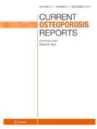
Following delivery, assessment should focus on basic life support, i.e. establishing airway, breathing, and circulation. In severe cases of OI, a small chest, pulmonary hypoplasia, intrinsic lung disease, airway collapse, and multiple fractures, potentially with flail segments, can all significantly impair an infant’s ability to effectively self-ventilate (Fig. 1). Pain from fractures is another important element.
Fig. 1
Plain radiograph of chest of an infant with severe osteogenesis imperfecta on a neonatal intensive care unit. There are multiple rib and vertebral fractures with some reduction in chest size. The infant was self-ventilating in low-flow oxygen at the time of the radiograph and did briefly show signs of respiratory failure on blood gas measurements but did not require continuous positive pressure support
Severe osteogenesis imperfecta itself is part of a spectrum of disease extending from lethal disease which is incompatible with life to infants with long bone deformities but no difficulties of self-ventilation. It is sometimes difficult to predict the precise severity and cause of disease based on antenatal evidence [3]. Thus, it is prudent to obtain a postnatal assessment by a clinical team experienced in OI at the earliest opportunity to help guide care. Indicators of severity relevant to the assessment of lethality include a small chest/pulmonary hypoplasia, “beading” of ribs, and marked shortening and angulation of long bones. Sadly, there are situations in which disease is so severe that the infant is unlikely to survive, even with the best care. In these situations, the usual ethical and legal principles apply. Clear and accurate information should be provided to the family where possible, with acknowledgement of uncertainties, together with an honest appraisal of the likelihood of various potential outcomes. Decisions should be made with close involvement of the family, in the best interests of the child.
Management of Respiratory FailureBoth the specialist team and any paediatric respiratory specialist involved in the care of infants with severe OI should have a good understanding of the multi-faceted pathophysiology of respiratory problems in OI. They should have a broad appreciation of the likely and potential long-term outcomes in severe OI. These are typically good from a neurocognitive perspective, though not always; one should recognise that the more severe the case, the more uncertain the outcome. In addition, it must be recognised that the literature is sometimes limited, with cases described as “lethal” which may or may not have been so with different care. Thus, there is risk that framing bias might lead to a self-fulfilling prophecy of lethality in some cases.
In situations where respiratory support is deemed necessary and appropriate, it is important to make sure that this is sufficient. There is a risk of undue caution allowing chronic under-ventilation, risking a vicious cycle of persistent collapse, recurrent chest infections, and gradual deterioration. In the context of severe OI, the need for respiratory support is most commonly for short-lived supplemental oxygen and/or non-invasive ventilation in the immediate days and weeks after birth. Effectiveness of non-invasive ventilation can be limited by specific difficulties such as achieving a good seal with a facemask; the weakness of bones and size of the fontanelle can mean that it is difficult to fit a facemask sufficiently well without the application of a degree of pressure that might be harmful (e.g. significant deformation of the skull vault). More prolonged supplemental oxygen and non-invasive positive-pressure ventilation may be required in some cases, and this may be necessary for months or a few years. In general, one can expect the need for ventilatory support to disappear or lessen over time as rib fractures heal, bones strengthen through the effects of bisphosphonate therapy, the chest grows, and the lungs mature. In extreme cases, adequate ventilatory support may require long-term invasive ventilation and tracheostomy. In such situations, one may anticipate this to be needed for many years.
Medical TherapyIn a newborn with severe OI, pain management is crucial. Some form of opiate analgesia is commonly required [4•]. With multiple fractures, a continuous opiate infusion may be the best approach in the first instance, although this can be weaned once the fractures stabilise over the course of a week or so.
Intravenous bisphosphonate treatment is effective in increasing lumbar spine bone mineral density, does not impair growth, and may reduce fracture rates and help preserve vertebral heights [1, 5,6,7,8]. Whilst trial data are limited, bisphosphonates have been used in infants with OI for more than 2 decades in specialist clinical centres across the world. Over that time, because of the cumulative experience of both their efficacy and safety, bisphosphonates have become standard of care for infants with severe OI. Rather than whether to treat, most commonly, the decisions are when, with which bisphosphonate, and how.
Bisphosphonates are an effective analgesic agent [4•]. Infants can be seen to settle after a first infusion of bisphosphonate, becoming more comfortable, with basic physical observations improving. However, whilst early treatment can be helpful, any decision to treat is, of course, a balance between benefit and risk. Timing of first infusions varies. One approach is to delay treatment for a few days where possible, until the usual physiological changes in the immediate postnatal period have been completed. First infusions of bisphosphonate are well known to cause acute phase reactions. In infants with severe OI, acute cardiovascular deterioration is well recognised as a potential serious adverse effect, perhaps particularly in those infants in whom there is already some cardiorespiratory compromise [9]. An infant under the care of one of the authors, who was undergoing intensive monitoring, was seen to develop significant pulmonary hypertension during a first infusion of pamidronate which resolved with cessation of the same. We take a cautious approach of admitting all infants with severe disease to a high dependency environment for their first infusion of bisphosphonate. To mitigate the risk of a severe acute phase reaction, we also ensure an infant is not vitamin D deficient and administer simple anti-pyretic medication, typically paracetamol, for several days after the first infusion.
Hypocalcaemia is a risk following first bisphosphonate infusions. Serum calcium levels drop during infusions and for a few days afterwards. Routine administration of calcium supplements for a few days following the first infusion usually avoids any significant problems. Symptomatic hypocalcaemia is very uncommon, but families should be warned of the risk and asked to get in touch if concerned (e.g. if the infant develops an intercurrent illness following admission which may increase the chance of problems, say gastroenteritis).
It is good practice to routinely counsel about risks and concerns regarding both short-term and long-term risks of bisphosphonate treatment in OI and to provide written information about the same [10]. This should be revisited at times beyond the immediate neonatal period, as the family’s understanding grows, and perspectives change.
There is no clear consensus on dose and frequency of administration of intravenous bisphosphonates. Annual doses of pamidronate have varied from 6 mg/kg to 12 mg/kg [6, 8]. It is common to start with more frequent and lower doses initially. We routinely use a starting dose of pamidronate of 0.5 mg/kg, typically administered over 4 h (Table 1). In exceptional cases, where the risk of cardiovascular instability is deemed high or the potential consequences of it are extreme, we have administered a starting dose of 0.25 mg/kg. Other similar regimens are used elsewhere, and zoledronic acid is employed by some.
Table 1 Example of a guideline for doses of pamidronate to be used in treating infants with severe osteogenesis imperfecta in the first year of life (after Senthilnathan [8])At the present time there are no other effective bone-targeted medical or cell-based therapies available for the treatment of infants with OI.
Vascular AccessAs bisphosphonate treatment is expected to be administered long-term, it is clear that venous access will be required both for blood sampling and drug administration. Peripheral venous access can be difficult in small infants and may get more difficult over time. As well as pain and distress, there are specific risks related to holding infants with OI for insertion of a peripheral catheter such as fracture. Central venous access devices (CVADs) largely avoid these risks, although insertion itself carries risks and there is the risk of serious sepsis, albeit that this is probably low with proper care [11•]. There have been cases of infants with OI experiencing frequent infections necessitating line removal, and it has been suggested that the impact of OI on soft tissues may predispose to problems with CVADs in small and severely affected infants. In truth, there is little published evidence to guide either routine use or avoidance of CVADs in OI, and some centres insert these routinely in all severely affected infants with good results. Other centres are more cautious, inserting CVADs either when clearly indicated on the balance of risk (e.g. difficulty inserting peripheral catheters which is likely to recur) or deferring routine insertion for a few months, thereby perhaps lessening the risks.
Orthopaedic ManagementInfants with severe OI are commonly born with significant long bone deformities, may have multiple long bone fractures, and limbs may lie fixed in extreme positions which may hinder care and handling (Fig. 2). Orthopaedic surgery is seldom required at this early stage. However, involvement of an orthopaedic surgeon is often helpful both to advise on management of limb fractures, perhaps to set the scene for future discussions about orthopaedic care, and/or to provide reassurance that nothing further needs to be done at that stage.
Fig. 2
Plain radiograph of left lower limb of the same infant with severe osteogenesis imperfecta shown in Fig. 1. There are fractures as well as shortening, widening, and angulation of the femur, tibia, and fibula. These skeletal features are indicative of generalised profound material weakness and low bone mass
Immediate management of fractures is generally conservative, with immobilisation, say with wool and crepe bandaging. Traction should not generally be used in infants with OI as it is often unnecessary and can result in harm such as further fractures.
FeedingNutrition is an essential element in the care of any infant. In critically ill infants with OI who are requiring some respiratory support, it may not be possible to feed them enterally. More commonly, the dilemma is whether they can be fed orally or require a nasogastric tube. In those unfamiliar with OI there is a tendency to perceive an infant with visible physical difference and fragile bones as requiring tube feeding, particularly in the high dependency setting. It is the role of the specialist team to ensure, where appropriate, that infants are fed orally, and mothers are helped to breast feed their child [2]. Often the practical instruction of mothers is undertaken by the clinical nurse specialist, who will also educate the neonatal nursing team and serve as a point of contact during the first few days and weeks as feeding is established. In severely affected infants on oral feeds, the need to manage them in a horizontal position to minimise vertebral compression fractures (see below) can present challenges, including with “winding”, for which we commonly use of preparations containing lactase or simethicone, as well as advising on safe physical methods.
Head ShapeThe head of an infant with severe OI may feel very soft due to both the quality of the skull bones and the number of Wormian bones. Together with excessive caution in handling and, sometimes, periods of high-dependency care, plagiocephaly is common and can lead to persistent marked deformity of the skull vault. Whilst the long-term effects of marked brachycephaly and other skull and cranio-cervical junction deformities in OI may be unclear, the potential risks are sufficient to justify attention to head shape in early infancy, i.e. at a time when intervention may have an effect. The potential risks of deformity include the effect on the anatomy of the cranio-cervical junction, including basilar invagination, and the presence in some cases of structural abnormalities of the cervical spine, e.g. severe cervical kyphosis [12•, 13]. Therefore, families should be shown how to vary the position of the infant’s head, and to manage any torticollis, to minimise the degree of deformity [2]. The role of aids such as specially designed pillows or helmets is still to be established [14, 15]. Whilst basilar invagination typically develops in later childhood, and both basilar invagination and significant cervical spine anomalies are rare, there may be a role for magnetic resonance scan imaging in infancy as a baseline and/or screening assessment.
Comments (0)